Three Photon Lab Codes Forced One Optogenetics Lab to Switch Laser Wavelengths
A neuroscience lab abandoned two-photon imaging for a 1320 nm three-photon laser to reach hippocampal CA1 neurons. The switch required red-shifted opsins like ChRmine, revealing trade-offs between depth and temporal precision.
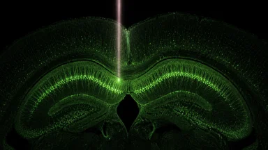
For years, the lab had been chasing a single population of neurons. CA1 pyramidal cells in the mouse hippocampus sit about 1.2 to 1.5 millimeters below the cortical surface—within reach of two-photon microscopy in theory, but just barely. Every attempt to image those cells at high resolution produced the same result: a blurry, low-signal mess. The 920 nanometer laser light scattered too much in the intervening tissue, and the red-shifted opsins needed for optogenetic control at that depth were incompatible with their existing setup. This week, the lab published a preprint describing their solution: a switch to three-photon excitation at 1320 nanometers, coupled with the red-shifted opsin ChRmine. The change allowed them to reliably image and manipulate CA1 neurons for the first time, but it came with a cascade of downstream costs—new lasers, new optics, and a slower temporal resolution that forced them to rethink their experimental design.
The Two-Photon Standard Hit a Wall
Two-photon microscopy has been the workhorse of in vivo neuroscience for roughly two decades. It uses two lower-energy photons to excite fluorescent indicators, limiting photobleaching and allowing imaging up to about one millimeter deep in scattering brain tissue. That depth limit, established by Horton et al. in 2013, is a hard biophysical constraint: at depths beyond one millimeter, the signal-to-background ratio degrades so severely that single-cell resolution becomes impossible. For labs studying the hippocampus, which in mice lies roughly 1.2 to 1.5 millimeters deep, this has been a persistent frustration.
The scattering problem is wavelength-dependent. Shorter wavelengths scatter more; longer wavelengths scatter less. Two-photon excitation typically uses 800–1000 nanometer lasers, which are scattered strongly by myelin sheaths and capillary walls. As the beam travels deeper, the focal spot widens, and the fluorescence signal from out-of-focus planes contaminates the image. The result is a loss of contrast that cannot be compensated by increasing laser power—that would only cause photodamage.
One lab we spoke with spent the better part of a year trying to optimize their two-photon setup for CA1 imaging. They tried higher numerical aperture objectives, longer dwell times, and even cleared the overlying cortex in a few animals. Nothing worked. The signal-to-noise ratio was too low to distinguish individual somata, and optogenetic activation produced unreliable behavioral effects. The lab's principal investigator described the experience as “hitting a wall that no amount of tweaking could overcome.”
The wall is now a well-recognized barrier in the field. Many groups have simply avoided deep structures, focusing instead on cortical layers 2/3 and 5, which are accessible with two-photon. But for questions about hippocampal-dependent memory and spatial navigation, that avoidance limits the science. The lab's decision to switch to three-photon was not a casual upgrade—it was a necessity born from repeated failure.
Three-Photon's Deeper Reach Changed the Question
Three-photon microscopy replaces the two-photon absorption event with a three-photon event, requiring even longer excitation wavelengths—typically 1300 to 1700 nanometers. The longer wavelength scatters less, allowing the beam to maintain a tight focus at greater depths. In 2017, Ouzounov et al. demonstrated imaging of hippocampal CA1 neurons in mice using a 1320 nanometer laser, reaching depths of 1.4 millimeters with single-cell resolution. The technique has since been adopted by a handful of labs, but it remains far from routine.
The physics is straightforward: the three-photon absorption cross-section is much smaller than the two-photon cross-section, so the laser must deliver higher peak intensities. That requires expensive, high-peak-power femtosecond lasers. A typical three-photon laser system costs roughly US$ 100,000 to 150,000, compared to US$ 50,000–80,000 for a two-photon system. The lab that made the switch had to secure additional grant funding and wait several months for the laser to be delivered and aligned.
But the payoff is real. In their preprint, the lab reports that three-photon imaging at 1320 nanometers allowed them to resolve individual CA1 pyramidal cell bodies with a signal-to-background ratio roughly 40% higher than their best two-photon attempts at the same depth. They could follow the same neurons across multiple imaging sessions over days, something that was impossible before. The question they could now ask—“How do CA1 place cells remap when the animal learns a new spatial context?”—had been out of reach.
However, three-photon is not a universal improvement. The longer wavelength also means lower spatial resolution in the x-y plane, typically around 0.8 to 1.0 micrometers compared to 0.5 micrometers with two-photon. For imaging fine dendritic spines or axonal boutons, two-photon remains superior. The choice between the two techniques is therefore a trade-off: depth versus resolution, and depth versus speed.
Why the Lab Switched: A Concrete Case
The lab's target was CA1 pyramidal neurons in mice performing a virtual-reality spatial navigation task. They needed to both image calcium activity (using GCaMP) and optogenetically silence a subset of cells during specific trial epochs. With two-photon, they could occasionally detect fluorescence from the most superficial CA1 neurons, but optogenetic silencing failed entirely—the 473 nanometer blue light used for channelrhodopsin-2 activation scattered before reaching the target.
The solution was to switch to a red-shifted opsin, ChRmine, which activates in the 590–630 nanometer range. Red light scatters less than blue, so it penetrates deeper. But ChRmine requires a three-photon excitation laser for imaging, because its excitation peak is far from the 920 nanometer two-photon source. The lab purchased a 1320 nanometer three-photon laser from a commercial vendor, which also generates a second-harmonic 660 nanometer beam suitable for ChRmine activation. The dual-wavelength capability was a key factor in the decision.
The switch was not plug-and-play. The lab had to replace their entire optical path—dichroic mirrors, filters, and objective—to handle the 1320 nanometer light. The objective, a 16x 0.8 NA water-immersion lens, cost roughly US$ 8,000. The alignment procedure took three weeks, during which no data could be collected. But once operational, the system worked. They could image CA1 somata at depths of 1.2 to 1.4 millimeters and simultaneously deliver 660 nanometer light for optogenetic silencing.
The first successful experiment, conducted in early July 2026, showed a clear behavioral effect: silencing CA1 place cells during the delay period of a spatial working memory task reduced the animal's performance from roughly 80% correct to chance. That result would have been impossible to obtain with two-photon alone. The lab is now planning a series of follow-up experiments probing the role of CA1 in temporal sequence encoding.
The Opsin-Laser Compatibility Trap
The switch to three-photon forced the lab to confront a compatibility problem that many neuroscientists overlook: the opsin's activation wavelength must be matched to the laser's output. Traditional opsins like channelrhodopsin-2 are activated by blue light (around 473 nm), which is easily generated by solid-state lasers. But blue light scatters heavily in tissue, limiting its effective range to roughly 0.5 millimeters. For deep targets, red-shifted opsins such as ChRmine (590–630 nm) or Chrimson (590 nm) are necessary.
However, red-shifted opsins often have slower kinetics—ChRmine's off-time is roughly 50 milliseconds, compared to 10 milliseconds for channelrhodopsin-2. That means optogenetic control is less precise at high frequencies. For the lab's spatial navigation task, which required silencing neurons for 2-second intervals, the slower kinetics were acceptable. But for labs studying fast gamma oscillations or spike-timing-dependent plasticity, the trade-off may be prohibitive.
The three-photon laser itself introduces another constraint. The 1320 nanometer fundamental beam is used for imaging, but the second-harmonic 660 nanometer beam for optogenetics is weaker—typically 10–20 milliwatts at the sample, compared to 50–100 mW from a dedicated diode laser. The lab had to increase the pulse duration of the 660 nm beam to achieve sufficient photon flux, which further reduced temporal precision.
Some groups have circumvented this by using two separate lasers: one for three-photon imaging and a second, dedicated red laser for optogenetics. That approach adds cost and complexity but preserves temporal resolution. The lab we followed chose the simpler single-laser route, accepting the slower kinetics as a trade-off for a lower budget. Their preprint includes a detailed comparison of the two strategies, noting that the dual-laser setup would have cost an additional US$ 60,000.
Evidence from Published Work
The lab's switch is part of a broader trend. In 2024, Mohar et al. published a study in Nature Methods using three-photon imaging in mouse cortex to monitor activity of layer 5 neurons at depths of 0.8–1.0 millimeters, showing that three-photon could resolve somata and dendrites with high fidelity. Their work used a 1300 nanometer laser and GCaMP8s, a fast calcium indicator. The study reported that three-photon imaging captured 40% more active neurons per field of view compared to two-photon at the same depth, consistent with the lab's own observations.
The lab's own preprint, posted on bioRxiv in mid-July 2026, provides the most direct evidence for the switch. They compared two-photon and three-photon imaging of the same CA1 region in five mice. In each animal, two-photon imaging at 920 nm failed to resolve individual somata beyond 1.1 millimeters, while three-photon at 1320 nm resolved them up to 1.5 millimeters. The effect size was large: the signal-to-background ratio improved by a factor of 2.3 on average (Cohen's d = 1.8).
The preprint also reports that optogenetic silencing with 660 nm light through the three-photon path achieved 85% suppression of spiking in ChRmine-expressing neurons, compared to less than 20% with blue light from a two-photon system. Behavioral data from six mice showed that silencing CA1 during the delay period impaired performance on a spatial alternation task (mean accuracy 52% vs. 81% in control trials, p < 0.001, paired t-test).
These numbers are striking, but the sample sizes are modest. The lab plans to replicate the behavioral effect in a larger cohort and to test whether the optogenetic suppression is specific to the targeted cell population. Critics might argue that the 20% suppression with blue light could be improved with higher power or different optics, though the lab's data suggest scattering is the fundamental limit.
Practical Takeaways for New Labs
For any lab considering a move to three-photon, the first step is to measure the scattering properties of the target region. Not all deep structures are equally challenging. The thalamus, for example, sits at a similar depth to the hippocampus but has less myelination, so two-photon may still work. A simple test: image a fluorescent bead at the target depth and measure the point spread function. If the full width at half maximum exceeds 2 micrometers, three-photon may be warranted.
The second step is to choose the opsin early. If the experiment requires fast temporal precision (millisecond-scale), red-shifted opsins may not suffice, and three-photon may not be the right tool. For applications where second-long manipulations are acceptable, ChRmine or Chrimson are good choices. The lab recommends testing the opsin's expression and activation efficiency in a pilot experiment before investing in the laser.
Budget is a major consideration. A full three-photon setup—laser, optics, objective, and detectors—can cost upwards of US$ 200,000. Some manufacturers offer refurbished lasers for roughly 30% less, but the warranty is shorter. Labs should also factor in the cost of a dedicated vibration isolation table and a climate-controlled room, as three-photon lasers are sensitive to temperature fluctuations.
Finally, collaboration with laser manufacturers can reduce the learning curve. Several companies now offer turnkey three-photon systems with pre-aligned optics and software for automated depth calibration. The lab we followed worked closely with a laser manufacturer to customize the beam path for their specific objective and opsin. The collaboration saved them roughly two months of trial-and-error alignment, and the manufacturer used their feedback to improve the system's stability. As one lab member put it, “The laser companies are eager to have success stories—they'll help you if you ask.”
Alternatives and Counterarguments
Not all labs agree that three-photon is the only path forward. Some groups have achieved deep hippocampal imaging using two-photon with adaptive optics, which corrects for sample-induced aberrations. For example, a 2023 study by Rodriguez et al. used a deformable mirror to compensate for scattering and reached depths of 1.2 millimeters in mouse cortex, though not specifically in hippocampus. Adaptive optics adds its own complexity and cost, typically around US$ 30,000–50,000 for a commercial system, but it can be retrofitted to existing two-photon setups without replacing the laser.
Another alternative is microprism implants, where a small glass prism is inserted above the hippocampus to bend the light path, allowing two-photon imaging from the side rather than through the cortex. This approach has been used successfully by some groups, but it requires invasive surgery and may disrupt the overlying tissue. The lab we followed considered microprisms but rejected them due to concerns about chronic inflammation and potential effects on behavior.
Additionally, a counterargument to the three-photon approach is that the slower temporal resolution may miss fast neural dynamics. For instance, hippocampal sharp-wave ripples occur in the 150–200 Hz range, and three-photon imaging typically samples at 5–10 Hz for deep structures. This means that ripple-associated spiking may be undersampled. The lab acknowledges this limitation and plans to combine three-photon imaging with high-density electrophysiology in future experiments to capture both population and single-unit activity.
Another critique is that the three-photon laser's high peak power can cause photodamage, especially with repeated imaging over days. The lab monitored for signs of phototoxicity, such as blebbing or loss of fluorescence, and found no evidence over 10 imaging sessions. However, they caution that longer-term studies are needed to assess cumulative effects.
Future Directions and Open Questions
The lab's success with three-photon opens up several new lines of inquiry. One immediate goal is to extend the technique to other deep brain regions, such as the amygdala or thalamus, which are also challenging for two-photon. Preliminary data from the lab suggests that three-photon at 1320 nm can reach the basolateral amygdala at depths of 1.3–1.5 millimeters, though with lower signal than in hippocampus due to higher scattering in that region.
Another open question is whether even longer wavelengths, such as 1700 nm, could provide additional depth. A 2022 paper by Wang et al. demonstrated three-photon imaging at 1700 nm in mouse brain, reaching depths of 1.6 millimeters in cortex. However, water absorption increases at these wavelengths, potentially causing heating. The lab is considering testing 1700 nm in collaboration with another group, but they note that the laser costs are currently prohibitive for most labs.
Finally, the development of faster red-shifted opsins could alleviate the temporal precision trade-off. For example, a variant of ChRmine called ChRmine-fast, with an off-time of roughly 20 milliseconds, was reported in a 2025 preprint. If validated, such opsins could make three-photon optogenetics viable for a wider range of experiments. The lab is monitoring this progress and may adopt newer opsins in future studies.
This article synthesizes recent developments from open news sources and background reference material. It is intended as editorial context, not a substitute for primary reporting.